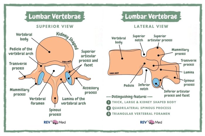
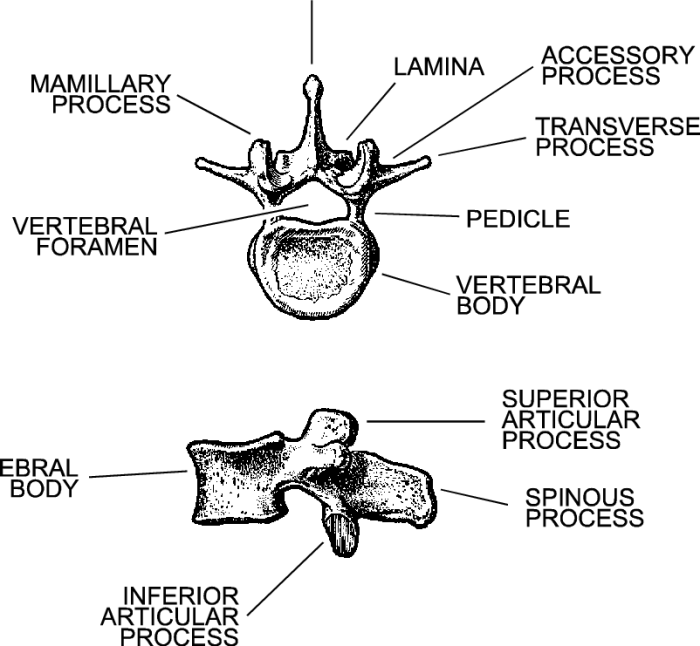
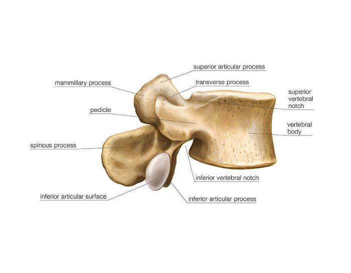
The lumbar vertebrae lateral view labeled provides a comprehensive visual representation of the lumbar spine, enabling healthcare professionals to assess its structure and identify potential abnormalities. This guide delves into the anatomy, imaging techniques, clinical significance, and radiographic interpretation of the lumbar vertebrae lateral view, equipping readers with a thorough understanding of this essential diagnostic tool.
Anatomy of the Lumbar Vertebrae: Lumbar Vertebrae Lateral View Labeled
The lumbar vertebrae are the five vertebrae located in the lower back, between the thoracic vertebrae above and the sacrum below. They are responsible for supporting the weight of the upper body and providing flexibility and mobility to the spine.
Each lumbar vertebra consists of a vertebral body, two pedicles, two laminae, and seven processes (two transverse processes, two superior articular processes, two inferior articular processes, and one spinous process). The vertebral body is the large, weight-bearing portion of the vertebra, while the pedicles and laminae form the vertebral arch.
The transverse processes extend laterally from the vertebral arch, while the superior and inferior articular processes project upward and downward, respectively, to articulate with adjacent vertebrae. The spinous process extends posteriorly from the vertebral arch and provides attachment for muscles and ligaments.
Imaging Techniques for Visualizing Lumbar Vertebrae

Lateral radiographs are the most common imaging technique used to visualize the lumbar vertebrae. They provide a two-dimensional image of the spine from the side, allowing for the assessment of spinal alignment, the detection of fractures, and the evaluation of degenerative changes.
Computed tomography (CT) and magnetic resonance imaging (MRI) are more advanced imaging techniques that can provide more detailed images of the lumbar vertebrae. CT scans use X-rays to create cross-sectional images of the spine, while MRI scans use magnetic fields and radio waves to create detailed images of the soft tissues surrounding the spine.
CT scans are particularly useful for visualizing bone structures, while MRI scans are better for visualizing soft tissues.
Clinical Significance of Lumbar Vertebrae Lateral View
Lateral view imaging of the lumbar vertebrae is essential for the diagnosis and management of a variety of spinal conditions. It can be used to assess spinal alignment, detect fractures, and evaluate degenerative changes. For example, lateral view imaging can be used to diagnose spondylolisthesis, a condition in which one vertebra slips forward on the vertebra below it.
It can also be used to detect spinal stenosis, a condition in which the spinal canal narrows, putting pressure on the spinal cord and nerves.
Radiographic Interpretation of Lumbar Vertebrae Lateral View

The normal radiographic findings of the lumbar vertebrae in lateral view include:
| Anatomical Structure | Radiographic Appearance |
|---|---|
| Vertebral body | Rectangular in shape, with a smooth, even surface |
| Pedicles | Thin, vertical columns of bone that connect the vertebral body to the vertebral arch |
| Laminae | Thin, flat plates of bone that form the roof of the vertebral arch |
| Transverse processes | Lateral projections from the vertebral arch that provide attachment for muscles and ligaments |
| Superior articular processes | Project upward from the vertebral arch and articulate with the inferior articular processes of the vertebra above |
| Inferior articular processes | Project downward from the vertebral arch and articulate with the superior articular processes of the vertebra below |
| Spinous process | A single, posteriorly projecting process from the vertebral arch that provides attachment for muscles and ligaments |
Common abnormalities that can be identified on lateral view images of the lumbar vertebrae include:
- Spondylolisthesis
- Spinal stenosis
- Herniated disc
- Fractures
- Degenerative changes
Advanced Imaging Techniques for Lumbar Vertebrae

Fluoroscopy is a real-time imaging technique that can be used to visualize the lumbar vertebrae in motion. It is particularly useful for assessing spinal alignment and detecting spinal instability.
Myelography is an imaging technique that uses a contrast agent to visualize the spinal canal and nerve roots. It is particularly useful for diagnosing conditions that affect the spinal cord and nerve roots, such as herniated discs and spinal stenosis.
Discography is an imaging technique that uses a contrast agent to visualize the intervertebral discs. It is particularly useful for diagnosing conditions that affect the intervertebral discs, such as degenerative disc disease and herniated discs.
Question Bank
What are the key anatomical features visible in the lumbar vertebrae lateral view?
The lateral view of the lumbar vertebrae showcases the vertebral body, pedicles, laminae, spinous process, transverse processes, and facet joints.
How does lateral view imaging help in evaluating spinal alignment?
Lateral view imaging allows for the assessment of spinal curvature, vertebral alignment, and the presence of any deformities or misalignments.
What are some common abnormalities that can be identified on lateral view images?
Common abnormalities include fractures, degenerative changes such as osteophytes and disc space narrowing, spondylolisthesis, and spinal stenosis.